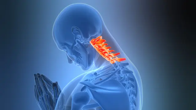
Cervical spinal stenosis occurs when the spinal canal or nerve exit openings in the neck narrow, placing pressure on the spinal cord or nerve roots. It is commonly caused by ageing, arthritis, injury, or degenerative spinal changes, leading to neck pain, stiffness, weakness, numbness, and reduced function. Degenerative changes affect most adults by age 50, making cervical stenosis increasingly common with age. At SpineMantra, specialised physiotherapy, posture correction, and targeted rehabilitation help reduce symptoms, improve mobility, and support long-term spinal health without invasive treatment whenever possible.
Why Choose SpineMantra?
- Our physiotherapists hold international MDT certification and specialise in non-surgical treatment of spine, ensuring safe and tailored care.
- We blend proven non‑surgical therapies with modern interventions tailored to patients’
- Our patient‑centric approach empowers you with self‑management tools.
- Conveniently located and committed to rapid, lasting relief, we prioritise your spine health.
- Bone Spurs: Prolonged wear and tear associated with arthritis or conditions such as Arthritis or age-related wear can cause bone overgrowth along the cervical spine. It narrows the spinal canal and compresses nearby nerves. These growths narrow the canal and press on nearby nerves.
- Herniated Disks: Disk herniation occurs when soft inner material bulges or leaks, pressing on the spinal cord or nerve roots.
- Thickened Ligaments: With age, the strong bands joining the spinal bones harden and broaden. They thicken and encroach into the space where nerves pass.
Traumatic Injuries: Serious incidents such as road traffic accidents or falls may cause fractures or dislocations. The bone fragments that follow form oedema or scar tissue that could irritate the spinal cord.
- Arm or Shoulder Pain: In cervical spinal canal narrowing, patients may experience shooting pain in the neck, shoulder, or arm with tingling or numbness.
- Hand Weakness: Cervical stenosis can weaken grip strength, reduce dexterity in hands, and cause difficulty holding everyday objects.
- Radiculopathy: Pain radiates from the site of compressive pathology with tingling, numbness, or weakness in the arms or legs, sometimes with reduced control.
- Myelopathy: When the spinal cord itself is compressed, one notices clumsiness and loss of balance along with a tingling sensation in both the arms and legs. It may also affect the bladder.
We at SpineMantra offer non-surgical cervical spinal canal treatment suited to individual needs. Our physiotherapy team uses posture correction, targeted exercises, manual therapy, and modalities such as ultrasound, heat therapy, and electrical stimulation to reduce pain and inflammation. When nerve irritation is severe, we may refer you to an ortho or neuro for medications. Our physiotherapists use evidence-based rehabilitation methods such as the MDT Technique for diagnosis, manual therapy, exercise therapy and electrotherapy.
If symptoms persist, we adjust rehab programs, apply spinal mobilisation and posture re-education techniques to ensure gradual improvement without invasive methods. It is a non-invasive procedure. We may also refer you to an orthopedic or neurologist in severe cases.
Dr. Saiantini Dey (PT) has strong expertise in spine and musculoskeletal physiotherapy. Our expert focuses solely on non-surgical approaches to treat cervical stenosis, helping patients avoid surgery through consistent physiotherapy-based management. Each procedure is tailored to the patient’s anatomy and lifestyle goals for optimal recovery.
- Symptom Review & Medical History: Our physiotherapists carefully assess your symptoms, posture, neck movements, and daily activity patterns to identify the cause of nerve compression. A detailed history of previous injuries, surgeries, and general health helps identify risk factors for the current cervical problem.
- Physical and Neurologic Examination: This involves checking your neck, arms and hands with sensory, motor, reflex, upper limb tension tests, hand function and coordination.
- X‑Rays: Routine X‑rays of the cervical spine provide a clear image of the alignment of the vertebrae and the presence of any bone spurs. They utilise a low dose of radiation to display where the spinal canal could have narrowed.
- MRI or CT Myelogram: MRI or X-rays are referred from diagnostic partners when needed, but our main focus remains clinical examination and movement assessment.