Why choose SpineMantra for spinal canal stenosis?
At SpineMantra, spinal canal stenosis is managed through specialised physiotherapy, personalised rehabilitation, movement-focused exercise programmes, and evidence-based pain management strategies. Our patient-centred approach helps reduce symptoms, improve mobility, restore function, and support an active, independent lifestyle with long-term spinal care.
- Age-Related Degeneration
As age catches up with people, the body naturally wears through the structures of the spine. Discs between vertebrae may dry up and decrease in size, joints can wear out and develop arthritis, ligaments may swell, and there is the potential to crush the space within the spinal canal.
- The Degenerative Disc Disease
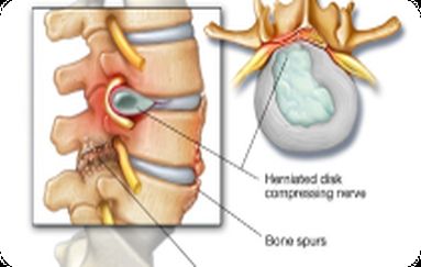
The spinal discs are cushioning disks that lose this cushioning effect as they wear out with age; as such, the vertebrae can move closer together. This may result in bone spurs and spinal canal stenosis, which will result in nerve compression.
- Bone spurs and arthritis
Due to osteoarthritis of the spine, the cartilage may wear out and cause the development of excess bone (bone spurs). These spurs can naturally jut into the spinal canal, where they reduce the space between the spinal cord and nerves.
- Thickened Ligaments
The spine has ligaments, which serve as the stabilisers; however, with time, ligaments may thicken and become rigid. This thickening may protrude into the space of the spinal canal, resulting in stenosis.
- Spinal Injuries
Fractures or dislocations in the spine may occur because of accidents, falls or sports injuries. Recovery of such injuries can cause overgrowth of bones or misalignments, and less space in the spinal canal.
- Tumours
There is the possibility of abnormal growths, benign and malignant, within the spinal canal. These projections can squeeze the spinal cord and nerve, resulting in symptoms of stenosis.
- Spinal Narrowing at Birth
Some individuals have a narrow natural canal. It is an inherited disorder which makes them more prone to show symptoms at an early stage of life, particularly when drugs are used with other degenerative alterations.
- Back or Neck Pain
There can be pain either continuously or on and off in the lower (lumbar stenosis) or neck (cervical stenosis) regions, and this pain usually exacerbates when one is on their feet or walking.
- Sharp shooting pain, Numbness or Tingling
Because of the nerve compression, pins and needles may appear in the arms, hands, legs or feet.
- Muscle Weakness
The limbs can become weak, resulting in difficulties while lifting objects, walking long distances or standing uniformly.
- Radiating Pain Sciatica-like Symptoms
The site, depending on which it affects, may be observed to radiate to the lower back, buttocks and legs or to the neck, shoulders and arms.
- Difficulty Walking, Standing
Others feel their legs heavy or cramped, usually when standing, which is relieved on sitting or bending forward.
- Disorganisation of order
The severe cases may hamper balance and coordination, causing an inclination to fall.
- Rare and Severe- Bowel or Bladder Dysfunction
Severe symptoms occur in instances where spinal nerves are compressed, and the result is the loss of control over the bladder or bowels that necessitates immediate medical care.
Spine Mantra will approach managing the spinal canal stenosis using advanced methods of physiotherapy combined with personalised care that will not involve unnecessary operations.
- Physiotherapy
We pay attention to specific exercises that reinforce muscles which hold the spine up, enhance flexibility and maintain the correct posture. This relieves pressure on the spinal cord and nerves, and you can move more freely and, luckily, with less pain.
- Pain Management
To aid in management, we can prescribe anti-inflammatory drugs, muscle relievers, or pain killers.
- Lifestyle Modifications
We advise you on how to maintain healthy weight levels, eliminate standing, and practice physical activities to unload your spine.
- Assistive Devices
Things like walking aids and supportive braces may help us improve our balance and stability, particularly in cases when we encounter weakness in our legs.
- Functional Limitation Scoring Symptom Profiling of Patients
We not only inquire as to the location of pain, but we also administer validated clinical assessment instruments such as the Oswestry Disability Index (ODI) to determine how the symptoms impact your daily life. This directive test would assist us in monitoring the improvement.
Data Insight: A study published in the Spine Journal (2019) indicates that more than 72 per cent of the patients with lumbar spinal stenosis have limitations in walking distance of below 500 meters without resting. Recognising this tolerance towards walking is a major element of early diagnosis.
- Dermatomal mapping or neurological examination should be done to find the cause of limb weakness.
Rather than a more generic physical exam, such as loss of sensation, weakness of reflexes, and muscle atrophy, we correlate them to the individual nerve roots. This indicates clearly the level of the spine that can be pressed.
Evidence shows that approximately 60 per cent of the cases of stenosis have multi-level entrapment of the nerves, a fact that necessitates the use of precision mapping in order to ensure that potential areas of a problem that are obscured are not overlooked.
- MRI with weight-bearing X-rays: Advanced Imaging Correlation
MRI should keep the status of a gold standard in confirming the spinal narrowing, and weight-bearing X-rays reflect the changes in the posture and position under load.
Data Insight: A 2021 meta-analysis reported that an MRI scan has a sensitivity of 94% to detect lumbar spinal stenosis, whilst functional images coupled with it enhance the quality of diagnosis when it comes to posture-related stenosis.
- McKenzie Mechanical Diagnosis & Therapy (MDT) Examination
Being the first clinic in entire Eastern India having a McKenzie-certified clinician, we implement MDT during the diagnosis period. This technique will involve making patients perform certain movements to confirm the diagnosis by noting the response of pain to those movements.
Why it Works: Clinical trials indicate that patients who centralise symptoms in the McKenzie assessment enjoy between 70 per cent and 80 per cent increased likelihood of having long-term positive outcomes without undergoing surgery.
- Nerve Conduction + EMG
In the case of a mismatch between symptoms and imaging results, electrodiagnostic tests are employed to establish the status and speed of the signal of the affected nerves.
Data Insight: According to the American Academy of Neurology, EMG may be useful in confirming nerve root involvement in more than 85 per cent of the suspected stenosis cases who exhibit borderline findings on MRI.
- Measurement of Cross-Sectional Area of Spinal Canal
We measure the diameter or cross-sectional area of the canal by MRI.
Data Insight: An area of less than 100 mm 2 indicates a stenotic canal, and an area of less than 75 mm 2 is normally related to severe symptoms such as neurogenic claudication, as recommended by the European Spine Journal.