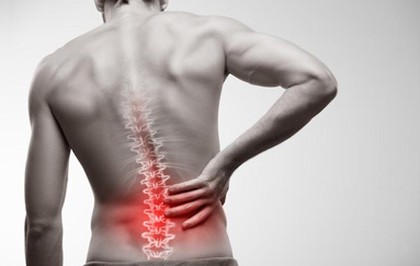
Lumbar canal stenosis is a degenerative condition where narrowing of the lower spinal canal compresses nerves supplying the legs. It commonly causes leg pain, numbness, heaviness, or weakness that worsens during standing or walking and improves with sitting or bending forward, known as neurogenic claudication. Age-related changes such as disc bulging, facet arthritis, and ligament thickening are common causes. At SpineMantra, treatment focuses on evidence-based, non-surgical physiotherapy to reduce nerve-related symptoms, improve mobility, restore function, and help patients regain independence through structured spine rehabilitation.
Who Is Most Affected by Lumbar Spinal Stenosis?
Lumbar spinal stenosis commonly affects adults over 50 due to age-related spinal degeneration. Risk factors include facet joint arthritis, ligament thickening, previous back injuries or surgery, repetitive spinal stress, family history of spinal disorders, and excess body weight, all of which can increase pressure on the lower spine and contribute to symptom progression.
Why Choose Spinemantra For Lumbar Spinal Stenosis?
At SpineMantra, lumbar spinal stenosis is managed through detailed clinical assessment and evidence-based physiotherapy. Treatment focuses on reducing nerve-related symptoms, improving mobility, and restoring function through personalised rehabilitation. Patient education and guided self-management help individuals understand symptom-modifying movements, participate actively in recovery, and manage symptoms confidently long term.
Lumbar spinal stenosis most commonly develops due to a combination of degenerative and structural changes that progressively reduce the space available for neural structures within the spinal canal.
Age-Related Degeneration (Lumbar Spondylosis)
With ageing, intervertebral discs lose hydration and height, facet joints undergo arthritic changes, and bone spur formation may occur. Along with gradual thickening of spinal ligaments, these changes collectively reduce the available space within the spinal canal and nerve root exit zones, contributing to symptom development over time.
Disc Bulge or Herniation
Degeneration or weakening of the intervertebral disc may allow disc material to bulge or protrude into the spinal canal. In an already narrowed canal, even modest disc bulging can increase nerve compression and provoke leg pain, numbness, or weakness, particularly during standing or walking.
Ligamentum Flavum Hypertrophy
The ligamentum flavum, which lines the posterior aspect of the spinal canal, may thicken and lose elasticity with age or repetitive loading. This inward buckling further narrows the canal, often worsening symptoms during spinal extension or prolonged upright postures.
Spondylolisthesis
Spondylolisthesis occurs when one vertebra slips forward relative to the one below it, commonly due to degenerative joint changes or structural defects. This vertebral displacement reduces canal and foraminal space, increasing mechanical compression of neural structures.
Congenital or Developmental Narrowing
Some individuals are born with a relatively narrow spinal canal due to anatomical variations such as shorter pedicles. While often asymptomatic earlier in life, these individuals have limited reserve space and may develop symptoms of stenosis earlier when age-related degenerative changes occur.
- Neurogenic Claudication
Neurogenic claudication is the hallmark symptom of lumbar spinal stenosis. Patients typically experience leg pain, heaviness, cramping, or fatigue that develops with standing or walking and progressively limits walking distance. Symptoms are characteristically relieved by sitting, bending forward, or leaning on support, such as a railing or shopping trolley.
- Leg Pain and Sensory Changes
Compression of spinal nerve roots may lead to pain radiating from the lower back into one or both legs. This pain may be sharp, burning, or electric in nature and often follows a nerve distribution. Patients may also report numbness, tingling, or altered sensation in the buttocks, thighs, legs, or feet, particularly after prolonged standing or walking.
- Lower Limb Weakness and Balance Difficulties
With ongoing nerve compression, some individuals develop weakness in the legs or feet, which may affect activities such as climbing stairs, rising from a chair, walking uphill, or maintaining balance. Reduced walking confidence and an increased risk of falls may be reported, especially in older adults.
Management of lumbar spinal stenosis at Spine Mantra is based on detailed clinical assessment and functional presentation rather than imaging findings alone. Treatment is non-surgical and focuses on symptom control, mobility optimisation, and functional independence, recognising that lumbar canal stenosis is often a predominantly non-mechanical condition.
Clinical Classification Using Mechanical Diagnosis and Therapy (MDT)
Mechanical Diagnosis and Therapy (MDT), also known as the McKenzie Method, is used primarily as a clinical assessment and diagnostic framework. MDT helps classify the nature of pain by analysing symptom response to repeated movements, sustained postures, and functional loading.
In lumbar spinal stenosis, MDT assessment assists in:
- Confirming non-mechanical or mixed pain behaviour
- Identifying symptom-modifying positions and postural biases
- Differentiating stenosis-related symptoms from other mechanical lumbar pathologies
- Guiding safe treatment direction and avoiding inappropriate mechanical loading
This step ensures that treatment is appropriately targeted and not based on generic exercise protocols.
Physiotherapy for Pain Modulation and Tolerance Building
Once lumbar canal stenosis is identified, physiotherapy focuses on reducing symptom irritability and improving tolerance to daily activities. Treatment aims to:
- Reduce nerve-related pain and leg symptoms
- Improve standing and walking tolerance
- Manage symptom flare-ups through graded exposure
- Address fear of movement and activity limitation
Rather than attempting to reverse structural narrowing, rehabilitation focuses on optimising function within available spinal capacity.
Mobility Restoration and Functional Conditioning
Restricted spinal and lower limb mobility often contributes to symptom aggravation in stenosis. Treatment includes controlled mobility work aimed at:
Improving lumbar and hip mobility within symptom limits
Enhancing pelvic and lower limb movement efficiency
Reducing excessive mechanical stress on neural structures during upright activities
Mobility is progressed cautiously, with continuous monitoring of symptom response.
Strength, Endurance, and Load Management
Reduced muscular endurance and poor load tolerance commonly worsen symptoms in lumbar spinal stenosis. Rehabilitation programs focus on:
- Improving trunk and lower limb strength
- Enhancing postural muscle endurance
- Building tolerance to prolonged standing and walking
- Gradual return to functional and occupational activities
Exercise prescription is individualised and adjusted based on symptom behaviour rather than fixed timelines.
Posture, Activity Modification, and Education
Patients are educated about posture, activity pacing, and daily movement strategies that minimise symptom aggravation. Emphasis is placed on:
Recognising symptom-relieving positions
Managing daily loads safely
Avoiding prolonged postures that increase neural compression
Developing long-term self-management strategies
Education plays a key role in reducing dependency on passive care and improving confidence in daily movement.
Non-Pharmacological Pain Management
As a physiotherapy-led clinic, Spine Mantra does not prescribe medications or administer injections. Pain management is addressed through active rehabilitation, movement-based strategies, graded exposure, and patient education aimed at improving tissue tolerance and reducing neural sensitivity over time.
Surgical Referral Pathway
Surgical intervention is considered only when conservative management fails or when progressive neurological deficits are identified. In such cases, appropriate referral is advised. Spine Mantra’s primary focus remains on maximising recovery and function through structured, evidence-based non-surgical physiotherapy.
Accurate diagnosis of lumbar spinal stenosis requires careful clinical reasoning, as symptoms often overlap with other spinal, neurological, and vascular conditions. At Spine Mantra, diagnosis is based on a structured combination of clinical history, physical examination, functional assessment, and imaging correlation.
Detailed Clinical History and Symptom Behaviour
A comprehensive history is taken to understand the exact nature, distribution, and progression of symptoms. Particular attention is given to posture- and activity-dependent symptom behaviour, such as worsening leg pain or heaviness with standing and walking and relief with sitting or forward bending. This pattern is characteristic of neurogenic claudication and helps differentiate lumbar canal stenosis from other causes of back or leg pain.
The duration of symptoms, changes in walking tolerance, night discomfort, and impact on daily activities are carefully documented to assess severity and functional limitation.
Comprehensive Physical and Neurological Examination
A detailed physical examination is performed to assess posture, spinal alignment, gait pattern, and functional mobility. Gait changes, balance confidence, and walking tolerance are observed. Neurological assessment includes evaluation of muscle strength, reflexes, and sensory changes in the lower limbs, looking for patterns consistent with nerve root involvement.
Spinal movement testing focuses on symptom response to different positions and movements. Reproduction or worsening of leg symptoms with lumbar extension and improvement with flexion provides important clinical evidence of dynamic canal narrowing affecting neural structures.
Imaging Correlation (When Indicated)
Magnetic Resonance Imaging (MRI) is recommended when clinically necessary to visualise the spinal canal, intervertebral discs, ligaments, and neural compression. Imaging findings are always interpreted in conjunction with clinical presentation and physical examination, rather than used in isolation.
When MRI is contraindicated, CT myelography may be considered to assess bony anatomy and canal dimensions. Plain radiographs may be used to evaluate spinal alignment, degenerative changes, or instability such as spondylolisthesis.
Exclusion of Differential Diagnoses
An essential part of diagnosis involves ruling out other conditions that can mimic lumbar spinal stenosis. These include peripheral vascular disease, peripheral neuropathy, hip pathology, and other spinal conditions such as disc herniation, infection, or tumours. Differentiation is based on symptom behaviour, clinical findings, and, where necessary, appropriate medical investigations.
Confirming that symptoms arise from structural canal narrowing — rather than vascular or peripheral neurological causes — ensures accurate diagnosis and guides appropriate treatment planning.